
What Do Dental X-Rays Show?
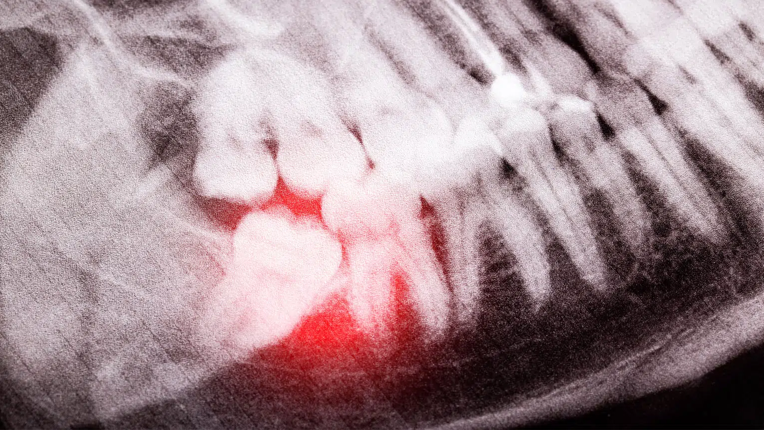




Dental X-rays, or radiographs, help dentists evaluate areas that cannot be fully seen during a visual exam. They may reveal decay between teeth, changes around roots, bone support, developing teeth, or the position of teeth below the gums.
Different images answer different questions
- Bitewings commonly show spaces between back teeth and bone levels.
- Periapical images show an entire tooth and nearby bone.
- Panoramic images provide a broad view of the jaws and teeth.
- Three-dimensional imaging may support selected surgical or diagnostic planning.
Timing should be individualized
The American Dental Association and U.S. Food and Drug Administration recommend selecting images after reviewing symptoms, oral health, age, disease risk, and prior records. A patient with new pain or active decay may need a different schedule from someone with stable health.
Recent images can sometimes be transferred when changing dentists, avoiding unnecessary repetition when they still answer the clinical question.
Ask why an image is recommended
Patients can ask what the dentist hopes to learn and how the result may affect care. Crown Point Smiles uses imaging as one part of a complete dental evaluation, not as a substitute for an examination.
How image selection is individualized
A dentist first reviews symptoms, current findings, prior images, decay and gum risk, age, and treatment history. An image should answer a clinical question. Bitewings, periapical images, panoramic images, and cone-beam computed tomography show different structures and expose different areas, so they are not interchangeable.
Understanding radiation and safety
Dental teams use protective equipment, appropriate positioning, and the smallest field that provides needed information. Pregnancy should be disclosed, but urgent diagnosis should not automatically be postponed. Patients can ask why an image is needed, whether a recent image can be transferred, and how the result may change care.
What an image cannot do
Radiographs do not replace a visual and tactile examination. Some cracks, early gum changes, soft-tissue conditions, and sources of pain may not appear clearly. Findings must be interpreted with symptoms and clinical tests.
Preparing for a useful conversation
Bring a current medicine list and describe what you have noticed, when it began, and what makes it better or worse. Mention relevant dental work, health conditions, tobacco use, dry mouth, pregnancy, anxiety, and previous complications. Clear background information helps Crown Point Smiles distinguish general education from what applies to your individual situation.
Questions patients can ask
- What findings support the diagnosis or recommendation?
- What happens if the area is monitored instead?
- What reasonable alternatives are available?
- What maintenance, limitations, and future costs should I expect?
- Which symptoms should prompt an earlier call?
Frequently asked questions
Do I need X-rays at every cleaning?
No universal schedule fits everyone. Timing is based on risk and current need.
Why might a three-dimensional scan be recommended?
It may help selected implant, surgical, airway, or diagnostic planning, but it is not routinely necessary for every patient.
Questions about dental X-rays
Ask what the image is intended to evaluate, whether an earlier image is still useful, and how the result could affect the treatment plan. X-rays are one part of an evaluation, not a substitute for an exam or a conversation about symptoms.
Timing should be individualized. A person with active decay, pain, gum disease, a recent injury, or extensive treatment history may need imaging at a different interval than someone with stable oral health.
Tell the dental team if you are or may be pregnant, have trouble tolerating sensors, or recently had images taken elsewhere. Those details help the team choose an appropriate view and avoid unnecessary duplication.
Questions worth asking
- What findings are most important in my case?
- What are the reasonable alternatives, including monitoring when appropriate?
- What should I expect during recovery or follow-up?
- Which symptoms should prompt me to call sooner?
Putting the guidance into practice
Images are selected to answer specific questions. Bitewing views may help reveal decay between teeth and assess bone levels, while other views may show roots, developing teeth, or a broader area of the jaws. The useful view depends on the concern.
When reviewing a recommendation, ask what cannot be seen during the visual exam, how the image could change care, and whether previous records can be compared. That turns radiation-safety principles into an individualized decision rather than a fixed calendar rule.
Before you decide
Make sure you understand the likely benefit, important limitations, expected timeline, and follow-up. Written instructions are useful when there are several steps. If symptoms or circumstances change, contact the dental office so the plan can be reassessed rather than assuming the original advice still applies.
Sources
- American Dental Association: X-Rays and Radiographs
- U.S. Food and Drug Administration: Dental Radiographic Examinations
Meet the Author
Dr. Mustafa Saad
Dr. Saad provides general dentistry with interests in oral surgery and dental implants, taking a calm approach and explaining treatment clearly.
Meet Dr. Saad